Anatomical Brain Drawing
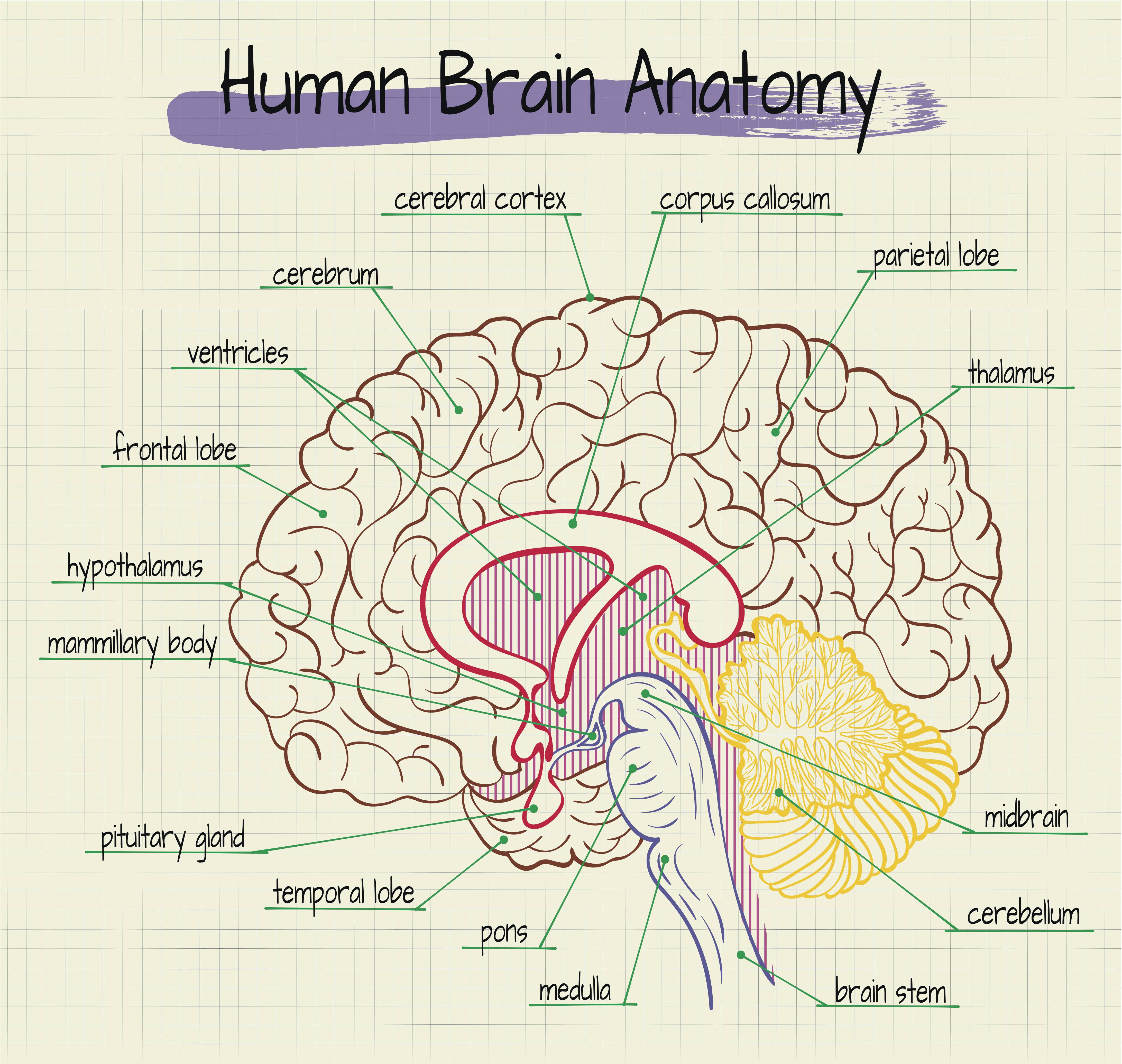
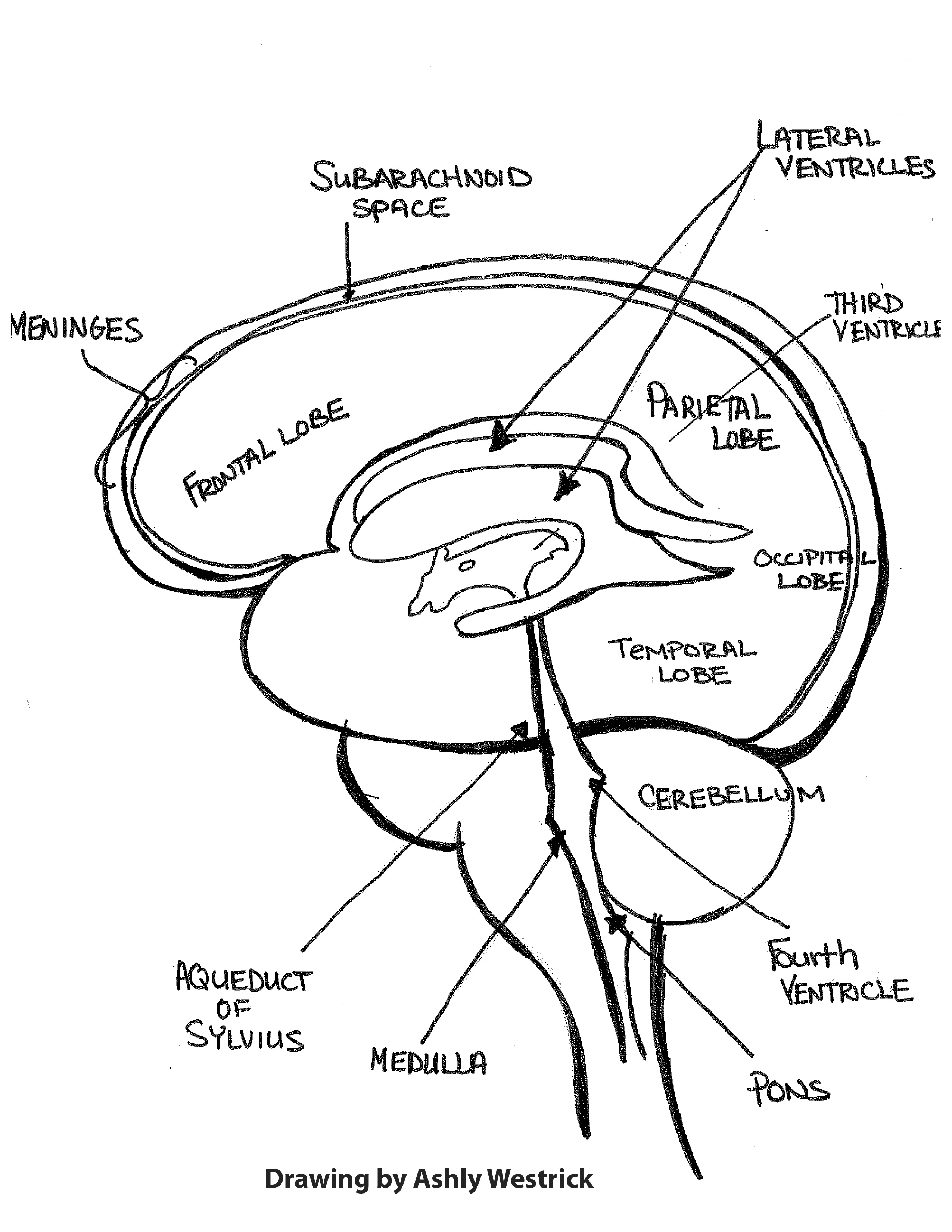
Anatomical Brain Drawing - The occipital lobe is the back part of the brain that is involved with vision. Web the midbrain is a part of the brain stem that connects the forebrain and the hindbrain. This fun and educational activity will help them develop their artistic skills and provide them with valuable knowledge about the brain's anatomical structures. Through easier tutorials, children will learn about the different parts of the brain. In intellectually superior mammals, such as humans, the cerebral cortex has protuberances. Web if you want more of a challenge, include anatomical parts, such as the brain stem. Try changing your search term. Web the parietal lobe houses wernicke’s area, which helps the brain understand spoken language. Unlock the art of creating a realistic brain drawing with authenticity and precision. The brain is one of the most fun parts of the body to draw. Forming the brain with a pencil sketch; Can you name these brain structures? Web anatomy of the brain. “a neurosurgeon’s overview of the brain’s anatomy” from the american association of neurological surgeons. Web table of contents. This neuroanatomical atlas is therefore perfectly adapted for the web. While drawing from imagination is a valuable skill, having reference images of the brain's anatomy can provide valuable guidance and ensure anatomical accuracy in your illustration. Outlining intricate parts of the brain; Cerebrovascular disease (stroke or brain attack): Woodcut blocks were used for the prints of figures in the vesalian anatomy. Figures of the brain appear to be done after external fixation in the work of willis. Web the parietal lobe houses wernicke’s area, which helps the brain understand spoken language. Web from “the beautiful brain: Web the midbrain is a part of the brain stem that connects the forebrain and the hindbrain. To draw an anatomically accurate brain, draw a. Can you name these brain structures? Use a combination of light and dark shading to add depth and. To draw an anatomically accurate brain, draw a curve in the shape of the lengthwise half of a. Web 18 internal brain anatomy resources. Sensory information is combined, evaluated, and compared to prior experiences, providing the brain with an accurate picture of. Through easier tutorials, children will learn about the different parts of the brain. Web the parietal lobe houses wernicke’s area, which helps the brain understand spoken language. It's not always easy remembering the parts of the brain. How many can you remember? Utilize anatomy books, online resources, or 3d models to study the structure of the brain. It's not always easy remembering the parts of the brain. Part of element ↖ back to the brain. Web the midbrain is a part of the brain stem that connects the forebrain and the hindbrain. Web an article in science daily on a research study about rem sleep and the pons, a part of the brain stem. It also reveals. Can you name these brain structures? Shading the center part of your brain drawing; The brain is one of the most fun parts of the body to draw. “a neurosurgeon’s overview of the brain’s anatomy” from the american association of neurological surgeons. Web the human brain is a complex organ, made up of several distinct parts, each responsible for different. Their illustrations, illustrators, and methods are discussed. It also reveals more structures. In intellectually superior mammals, such as humans, the cerebral cortex has protuberances. Use a combination of light and dark shading to add depth and. The drawings of santiago ramón y cajal” at grey art gallery, “tumor cells of the covering membranes of the brain,” from 1890. The brain is an organ made up of neural tissue. Shading the center part of your brain drawing; Through easier tutorials, children will learn about the different parts of the brain. The brainstem connects the brain. Try changing your search term. Woodcut blocks were used for the prints of figures in the vesalian anatomy. Try changing your search term. It's not always easy remembering the parts of the brain. Figures of the brain appear to be done after external fixation in the work of willis. Learn techniques to capture the intricate details and textures of the brain's anatomy. The association areas also work to develop plans of action that are sent to the brain's motor regions in order to produce a change in the body through muscles or glands. Scroll through the images with detailed labeling using our interactive interface. Web the human brain is a complex organ, made up of several distinct parts, each responsible for different. Web the anatomy of the human brain it is characterized by the following parts: Unlock the art of creating a realistic brain drawing with authenticity and precision. Sensory information is combined, evaluated, and compared to prior experiences, providing the brain with an accurate picture of its conditions. Web an average adult brain weighs 3 pounds and is composed of 60%. The occipital lobe is the back part of the brain that is involved with vision. The brainstem connects the brain. While drawing from imagination is a valuable skill, having reference images of the brain's anatomy can provide valuable guidance and ensure anatomical accuracy in your illustration. This neuroanatomical atlas is therefore perfectly adapted for the web. It's not always easy remembering the parts of the brain. It also reveals more structures. Use a combination of light and dark shading to add depth and. Web explore the structure and function of the human brain in 3d, with interactive models, videos, and articles on brainfacts.org. Web from “the beautiful brain: The drawings of santiago ramón y cajal” at grey art gallery, “tumor cells of the covering membranes of the brain,” from 1890. Cerebrovascular disease (stroke or brain attack): Woodcut blocks were used for the prints of figures in the vesalian anatomy. Web the human brain is a complex organ, made up of several distinct parts, each responsible for different functions. Web axial mri atlas of the brain. It is involved in many functions, such as vision, hearing, movement, and arousal. Cerebral cortex is a tissue layer that forms the brain’s outer covering, whose thickness fluctuates from 2 to 6 millimeters.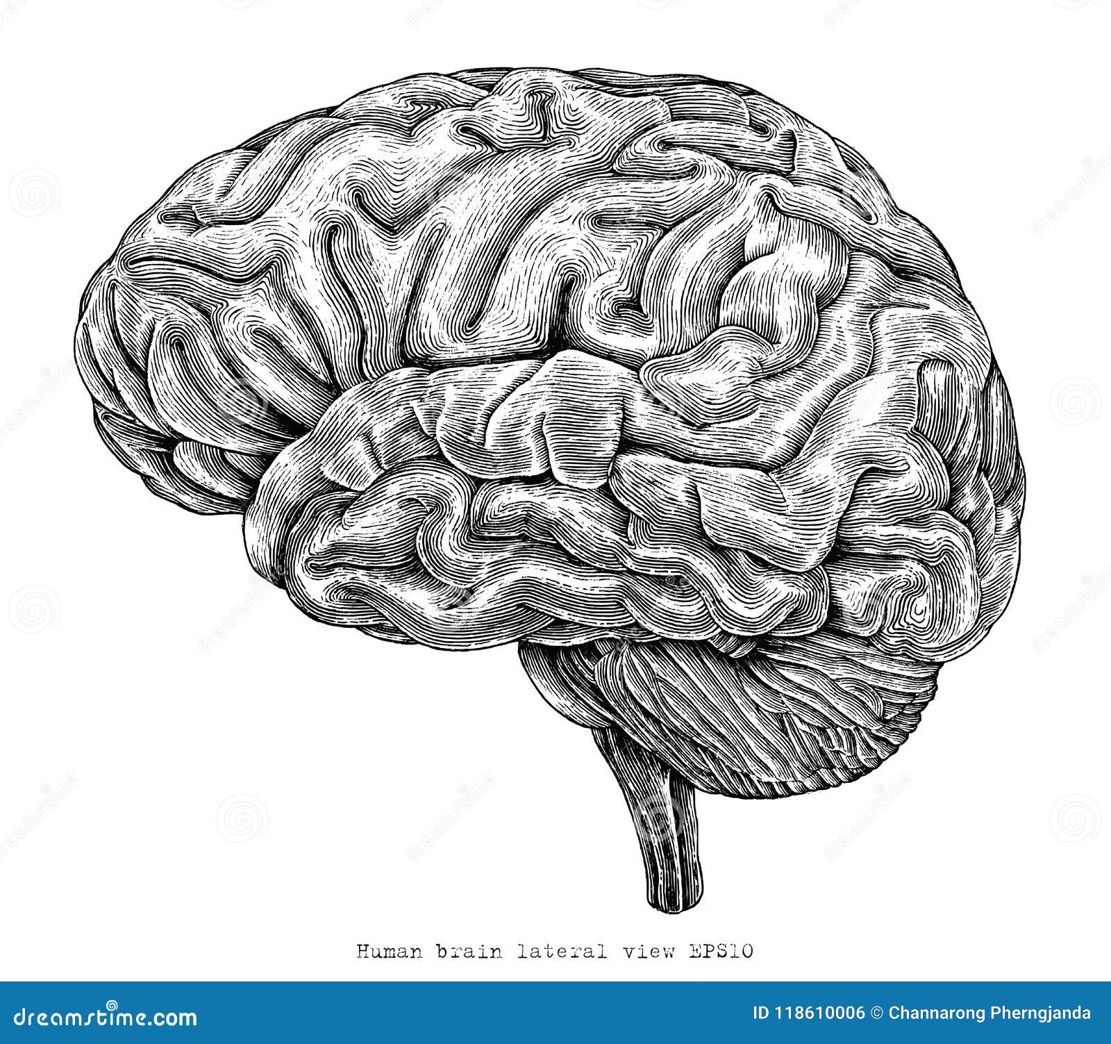
Human Brain Lateral View Hand Drawing Vintage Engraving Illustration
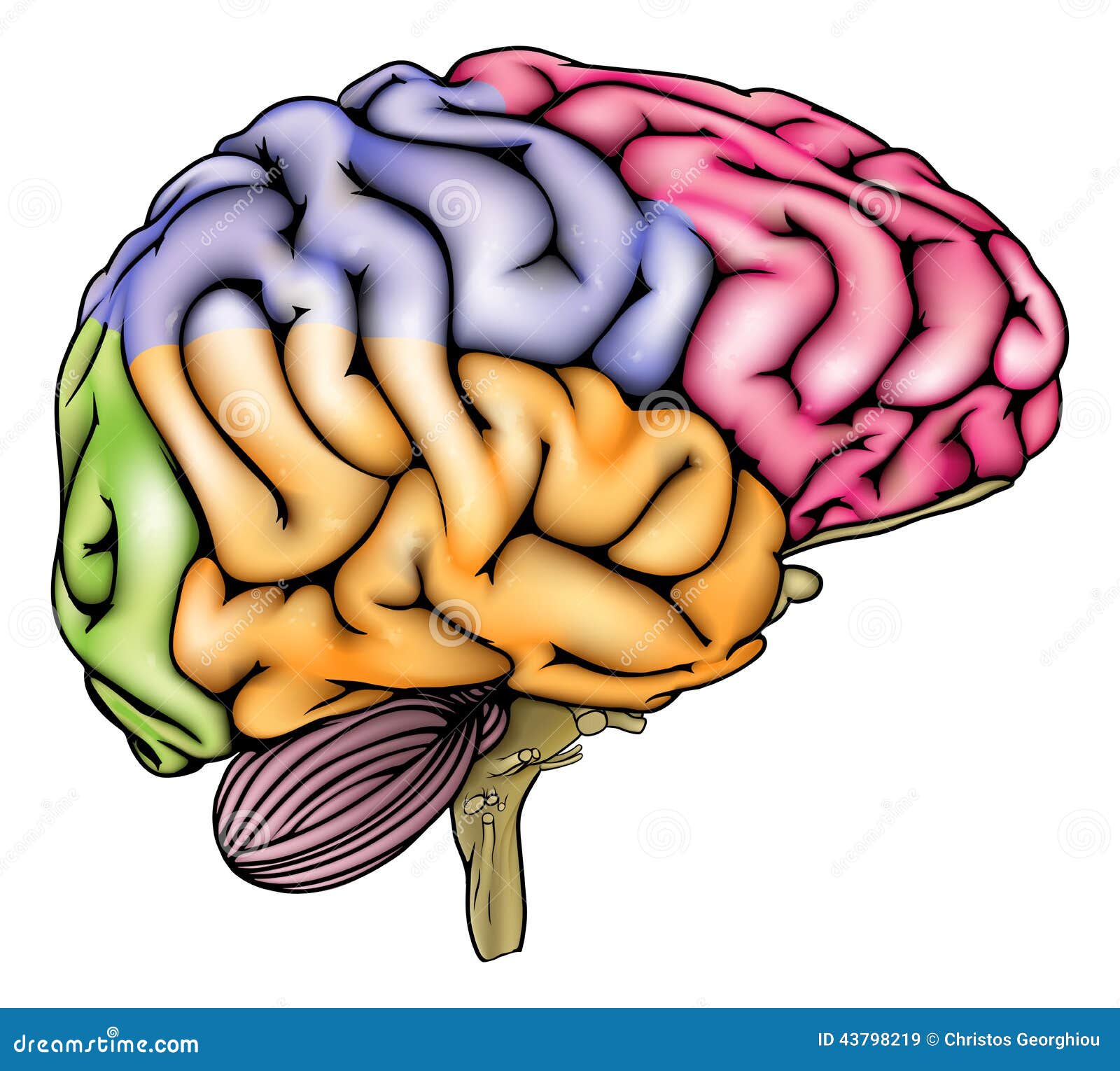
Human Brain Anatomy Sectioned Stock Vector Illustration of isolated
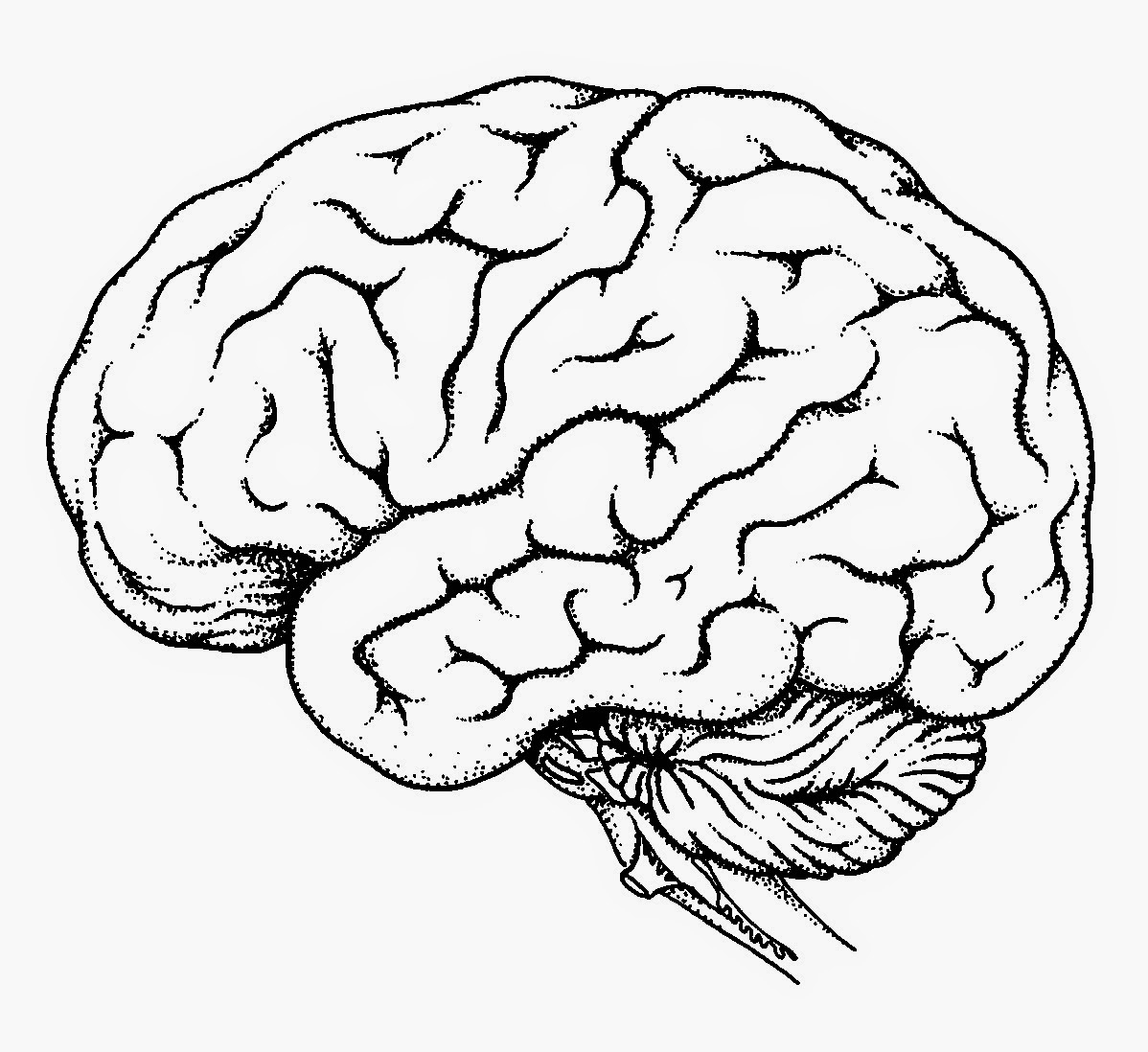
Human Brain Drawing at GetDrawings Free download

Scientific Illustration Brain anatomy, Medical anatomy, Anatomy

Human Brain Anatomy Stock Vector Image 44353466
brainanatomy Rachel Gold

How to Draw a Brain 14 Steps wikiHow
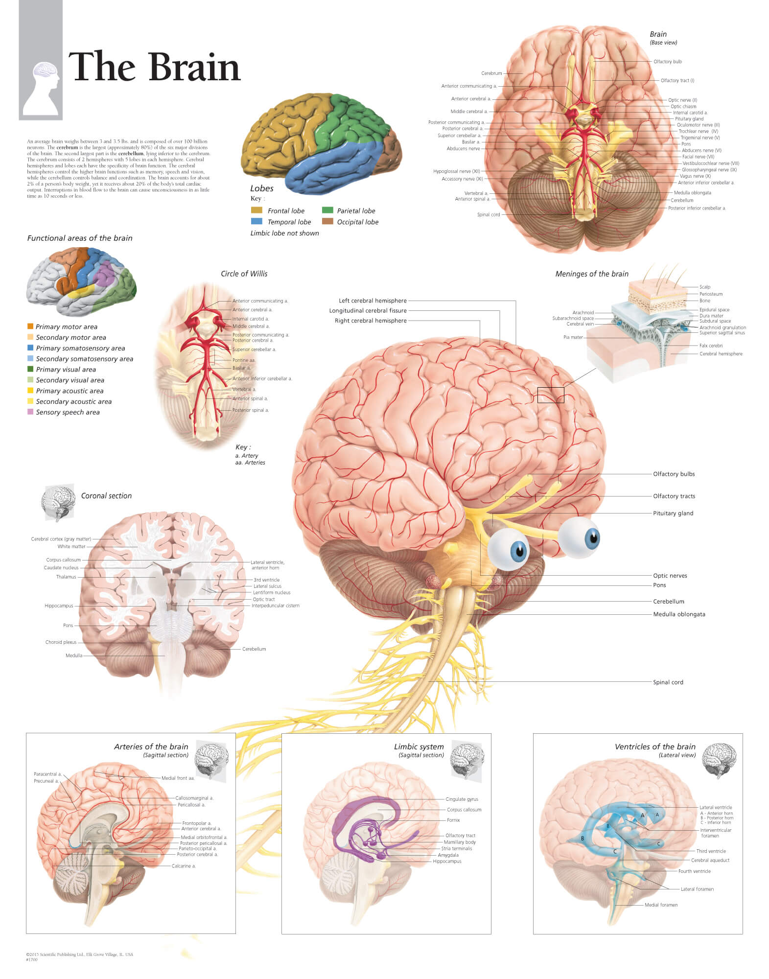
The Brain Scientific Publishing
Brain 101 An Overview of the Anatomy and Physiology of the Brain
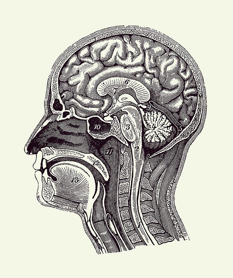
Normal Anatomy of Human Brain Vintage Print 2 Drawing by Vintage
Scroll Through The Images With Detailed Labeling Using Our Interactive Interface.
In Intellectually Superior Mammals, Such As Humans, The Cerebral Cortex Has Protuberances.
Web An Article In Science Daily On A Research Study About Rem Sleep And The Pons, A Part Of The Brain Stem.
Perfect For Clinicians, Radiologists And Residents Reading Brain Mri Studies.
Related Post: